
Building a Mac-Based Diagnostic Imaging Workstation: Display Calibration, Viewer Validation and Quality Assurance
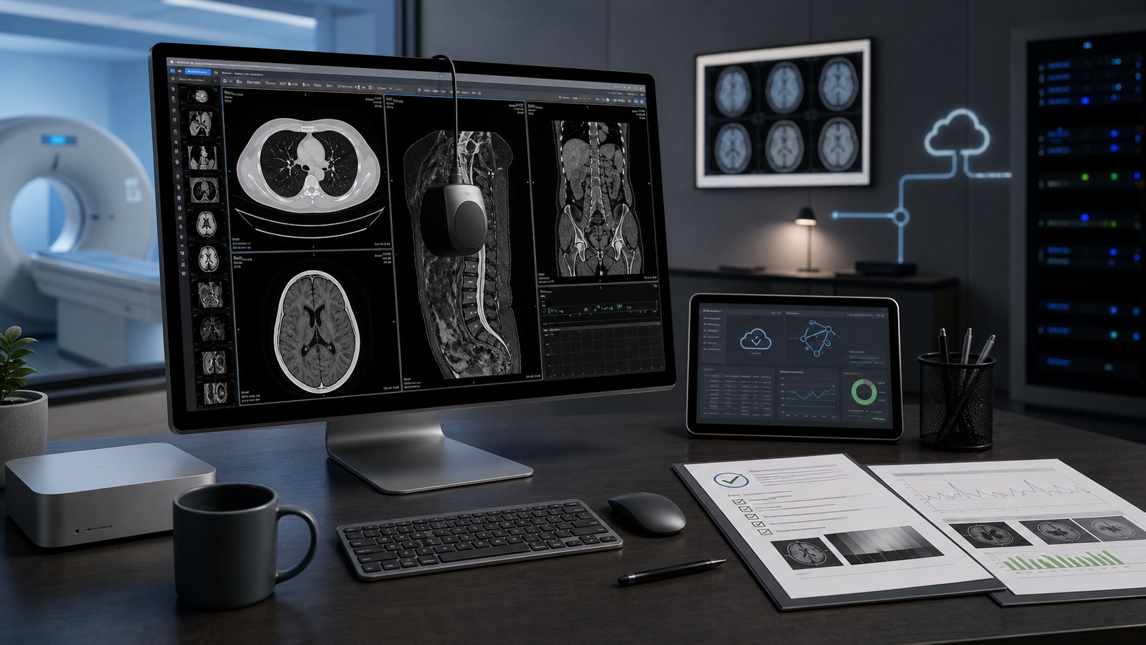
A dependable Mac imaging workstation emerges only when the computer, display, DICOM viewer, network, viewing environment and quality-assurance process are designed and tested as one clinical system.
In this article
A workstation is a system, not a single product
A private clinic purchases a high-specification Mac and premium display, installs a DICOM viewer and assumes the reporting station is ready. The images look sharp. Yet nobody has documented the display settings, measured the room lighting, checked measurement accuracy or observed how the viewer behaves when the PACS returns incomplete or compressed data.
That is powerful hardware, not yet a validated Mac diagnostic imaging workstation. Reliability depends on the presentation chain: hardware, display, software, connectivity, environment, configuration, users and quality controls. Visual quality alone is not evidence that the system is suitable for its intended clinical task.[4][5]
Each part contributes something different to the final clinical system:
Powerful Mac
Processing, graphics throughput and responsiveness.
High-quality display
Resolution, luminance potential and viewing geometry.
Calibrated display
A measured, controlled grayscale response.
Validated DICOM viewer
Tested image handling and clinical functions.
Locally validated workstation
Evidence that the complete chain works in the local environment.
Quality-controlled workflow
Confidence that performance remains acceptable over time.
Start with the intended clinical use
Requirements should begin with one question: what decisions will this workstation support? Primary interpretation, secondary review, procedural planning, emergency consultation, teaching, research and multidisciplinary meetings need not share identical configurations.
Define the modalities, users, location, workload, processing tools, display class, network dependencies and local quality-assurance obligations. A system suitable for routine CT, MR or radiography review cannot automatically be assumed suitable for every specialised application. Intended use should drive the evidence required, not the prestige of the hardware.[4][5]
Choose Mac hardware for the workload
Select the Mac around representative clinical studies. Processor and graphics capability affect scrolling, multiplanar reconstruction and three-dimensional visualisation. Memory influences how many large studies remain active. Fast storage supports caching and import, while network performance often determines how quickly remote studies become usable.
Check display outputs, macOS and viewer compatibility, sustained thermal behaviour, backup strategy and expected support life. Standard CT and MR review may have different needs from very large volumetric datasets, advanced 3D work or multi-display workflows. Confirm Falcon MD’s current system requirements and test the proposed configuration with the organisation’s real workload.[9]
The display is part of the clinical chain
Resolution and brightness matter, but diagnostic review also depends on physical size, pixel density, minimum and maximum luminance, luminance ratio, grayscale response, uniformity, reflections, pixel defects, viewing angle and ageing. AAPM guidance therefore treats display quality assurance as a programme rather than a one-time brightness check.[2][4]
DICOM Part 14 defines the Grayscale Standard Display Function, or DICOM GSDF. It specifies a predictable relationship between presentation values and visible luminance, reflecting the human visual system’s changing sensitivity across dark and bright levels. DICOM display calibration aims to make grayscale contrast steps more consistently perceptible; it does not make all monitors equivalent or guarantee equal information content.[1]
A preset labelled “DICOM” is not necessarily measured radiology monitor calibration. Calibration should use an appropriate sensor, observe any specified warm-up time, record the result, account for ambient light and be followed by visual test-pattern checks and objective measurements where required. Periodic reassessment is needed as displays age or configurations change. Apple’s guidance for its medical-imaging reference modes states that diagnostic use requires calibration with its medical-imaging calibrator and a compatible DICOM viewer, showing why a preset alone is insufficient.[2][3]
Control the viewing environment
Ambient light reflected from the screen raises the effective black level and can hide low-contrast detail, especially in darker regions. Conversely, a completely dark room is not always the most comfortable environment. The objective is controlled, repeatable lighting that is included in display assessment.[1][2][6]
Position the workstation away from windows and direct light, remove glare, keep the screen clean and maintain similar conditions between calibration and clinical use. Consider desk lighting, bright secondary monitors, user posture and fatigue. Even a calibrated display can underperform when its environment changes substantially.[2]
Validate the DICOM viewer clinicians will use
Falcon MD is the central viewing environment in this Mac radiology workstation. Official documentation describes 2D review, window and level, zoom and pan, measurements and annotations, multiplanar and curved reconstruction, volume rendering, series synchronisation, study comparison, multi-window working and PACS integration.[9]
Validation should focus on the functions the organisation relies on. With representative local studies, verify patient, study and series loading; image order and orientation; window and level; zoom, pan and cine; distances, angles and regions of interest; annotations; MPR, fusion and 3D behaviour; priors; metadata; exports; compression; error messages; and study completeness.[5][8]
Check measurements against known dimensions or a suitable test object. Include edge cases such as multiframe objects, oblique acquisitions, mixed series, non-ASCII names, large studies and interrupted imports. A feature list demonstrates capability; local testing demonstrates suitability.[5][8]
Validate PACS, DICOM and cloud connectivity
Test Falcon MD against the exact PACS, routers, gateways and cloud services in use. Include query/retrieve, storage, permissions, transfer syntaxes, character sets, large-study performance, duplicate handling, reconciliation, interrupted transfers and recovery. DICOM and IHE specifications guide integration, but the route still needs local testing.[5][7][13]
Falcon MD publishes a DICOM Conformance Statement documenting its declared storage, query/retrieve and DICOMweb behaviour. It is essential integration evidence, but not a substitute for site testing. DICOM Part 2 explains that comparing conformance statements helps determine what communication might be supported and calls for test procedures with specific equipment. Record successful paths and known limitations.[7][10]
Where available, Falcon Cloud can make studies accessible across Mac, iPad and iPhone and support continuity between locations. Assess it as a defined cloud-access layer, not as an assumed replacement for every PACS, archive or governance function. Availability is still expanding, so regional access and operational arrangements should be confirmed.[11]
Illustrative case: a private radiology workstation
A radiologist in a private clinic initially chooses a powerful Mac and premium display mainly for resolution and brightness. The team then defines the CT, MR and radiography workload; calibrates the display; records room lighting; configures Falcon MD; verifies measurements; tests representative studies and priors; validates PACS operations; and documents backup and recovery.
A controlled transfer test reveals that an older PACS node does not negotiate a required compressed transfer syntax as expected. The route is reconfigured and retested before routine use. Hardware specifications alone could not reveal this local workflow problem.
Illustrative case: workstation review with mobile continuity
A surgeon performs detailed CT and MR review and planning in Falcon MD on a configured Mac. Selected studies are available through Falcon Cloud where deployed. Away from the workstation, Falcon Mx on iPad or iPhone can support mobile access: checking whether a study has arrived, reviewing a relevant series, discussing a case or deciding that fuller Falcon MD review is required.[9][11][12]
The controlled workstation remains the environment for work that depends on its validated display, tools and procedures. Mobile access is useful when it supports the workflow around that workstation rather than replacing the validation decisions made for it.[12]
Quality assurance does not end after installation
Validation is a controlled baseline, not a permanent certificate. Reassess relevant parts after macOS or Falcon MD updates, display recalibration, hardware replacement, PACS or network changes, security updates, new modalities, new compression methods, user-configuration changes or incidents. Software-validation principles similarly link assurance to intended use, documented evidence and change control.[2][4][8]
Keep a simple record of hardware and software versions, calibration reports, ambient conditions, test studies, measurement and connectivity results, known limitations, corrective actions, dates and responsible personnel. Train users in approved configurations, error escalation, downtime procedures and the distinction between workstation review and mobile access.[4][5][8]
Workstation validation checklist
The retained evidence should demonstrate that the complete imaging chain, not just individual components, was assessed for its intended use.[1][2][4][5][7][8]
- 01
Document whether the workstation supports primary interpretation, secondary review, planning, teaching, research or another defined purpose.
- 02
List supported modalities, specialised applications and any excluded workflows.
- 03
Record processor, graphics, memory, storage, display outputs, network interfaces and thermal performance.
- 04
Record the macOS version, Falcon MD version and approved update policy.[9]
- 05
Verify resolution, physical size, luminance range, uniformity, pixel condition and viewing geometry.
- 06
Calibrate with a suitable sensor, retain the report and document warm-up and measurement conditions.
- 07
Measure or otherwise assess room lighting, reflections, glare and nearby light sources.
- 08
Record layouts, tools, viewer preferences, PACS nodes and user-specific settings.
- 09
Test normal studies, large datasets, multiframe objects, unusual orientations and relevant edge cases.
- 10
Check distance, angle and region-of-interest measurements against known values.
- 11
Verify query/retrieve, storage, study completeness, transfer syntaxes, character sets and permissions.[7][10]
- 12
Test interrupted transfers, duplicates, unavailable nodes, incomplete studies and clear user error messages.
- 13
Confirm Falcon Cloud availability and define when Falcon Mx may be used for mobile access around the workstation workflow.[11][12]
- 14
Verify user accounts, privileges, authentication, session handling and approved data-export routes.
- 15
Document local cache, archive, recovery and downtime procedures.
- 16
Define when macOS, Falcon MD, PACS, network, display or hardware changes trigger partial or full revalidation.
- 17
Schedule display checks, recalibration, performance review, incident review and user refresher training.
Conclusion
Building an Apple medical imaging workstation involves more than premium hardware. A dependable system combines an appropriately specified Mac, a suitable and calibrated display, Falcon MD as the DICOM viewer for Mac, tested PACS or cloud connectivity, controlled lighting, documented DICOM viewer validation, user procedures and continuing medical imaging quality assurance.
Falcon Cloud can provide a cloud access layer where available, while Falcon Mx extends access to iPad and iPhone without replacing the full workstation. A well-designed Mac imaging workstation is not defined by a single specification. It is defined by how reliably the complete system supports the clinician’s intended work.[9][11][12]
Frequently asked questions
These questions commonly arise when a clinic turns a Mac setup into a governed imaging workstation:
Does a high-resolution Mac display automatically create a diagnostic workstation?
No. Resolution is only one property of the display, and the display is only one component of the imaging chain. The monitor, DICOM viewer, room lighting, measurements, PACS connectivity, user configuration and QA process must all be suitable and locally validated.[2][4][5]
Is selecting a DICOM display preset the same as calibrating the monitor?
No. A preset selects predefined operating characteristics. Calibration measures the display’s actual response, applies or verifies the required correction and produces evidence that performance is acceptable under the intended viewing conditions. Apple’s medical-imaging guidance also distinguishes preset selection from measured calibration.[2][3]
Does a DICOM Conformance Statement guarantee PACS interoperability?
No. It documents the services, roles, object classes and transfer mechanisms declared by an implementation. Comparing statements helps predict compatibility, but DICOM Part 2 identifies real testing with specific equipment as part of interoperability validation.[7][10]
How often should a display be recalibrated or the workstation revalidated?
The interval should follow the display manufacturer’s instructions, local medical-imaging quality-assurance programme and applicable professional requirements. Additional checks should follow significant software, hardware, PACS, network or environmental changes rather than waiting for the next scheduled review.[2][4][8]
Where does mobile access fit in a Mac workstation workflow?
Mobile access can support continuity around the workstation, such as checking study availability, reviewing a relevant series or discussing a case. Work that depends on calibrated display conditions, validated tools and local reporting procedures should remain tied to the configured workstation.[9][12]
References
Sources cited in this article
- [1]DICOM PS3.14: Grayscale Standard Display Function
DICOM Standards Committee / NEMA
DICOM grayscale display function standard.
- [2]Display Quality Assurance
American Association of Physicists in Medicine
AAPM Report 270 on display quality assurance.
- [3]Studio Display XDR Technology Overview
Apple Inc.
Apple documentation for Studio Display XDR technology and medical imaging reference modes.
- [4]ACR-AAPM Technical Standard for Diagnostic Interpretation Displays
American College of Radiology
Technical standard for diagnostic interpretation displays.
- [5]ACR-AAPM-SIIM Technical Standard for Electronic Practice of Medical Imaging
American College of Radiology / AAPM / SIIM
Technical standard for electronic medical imaging practice.
- [6]The effects of ambient lighting in chest radiology reading rooms
Journal of Digital Imaging / PubMed
Study of ambient lighting effects in radiology reading rooms.
- [7]DICOM PS3.2: Conformance
DICOM Standards Committee / NEMA
DICOM conformance statement and interoperability documentation.
- [8]General Principles of Software Validation
U.S. Food and Drug Administration
FDA guidance on software validation principles.
- [9]
- [10]Falcon MD DICOM Conformance Statement
iCat Solutions Ltd.
Falcon MD DICOM conformance statement, revision 1.1.
- [11]Falcon Cloud: Continuity Beyond the Workstation
iCat Solutions Ltd.
Falcon Cloud product documentation.
- [12]
- [13]