
The Diagnostic Viewer in Veterinary Imaging: Preserving Study Integrity from Acquisition to Consultation
An imaging examination is more than a collection of pictures. Its diagnostic value depends on whether the images, series, metadata and clinical context remain intact as the study moves between acquisition, review and consultation.
In this article
A DICOM study is not simply a folder of images
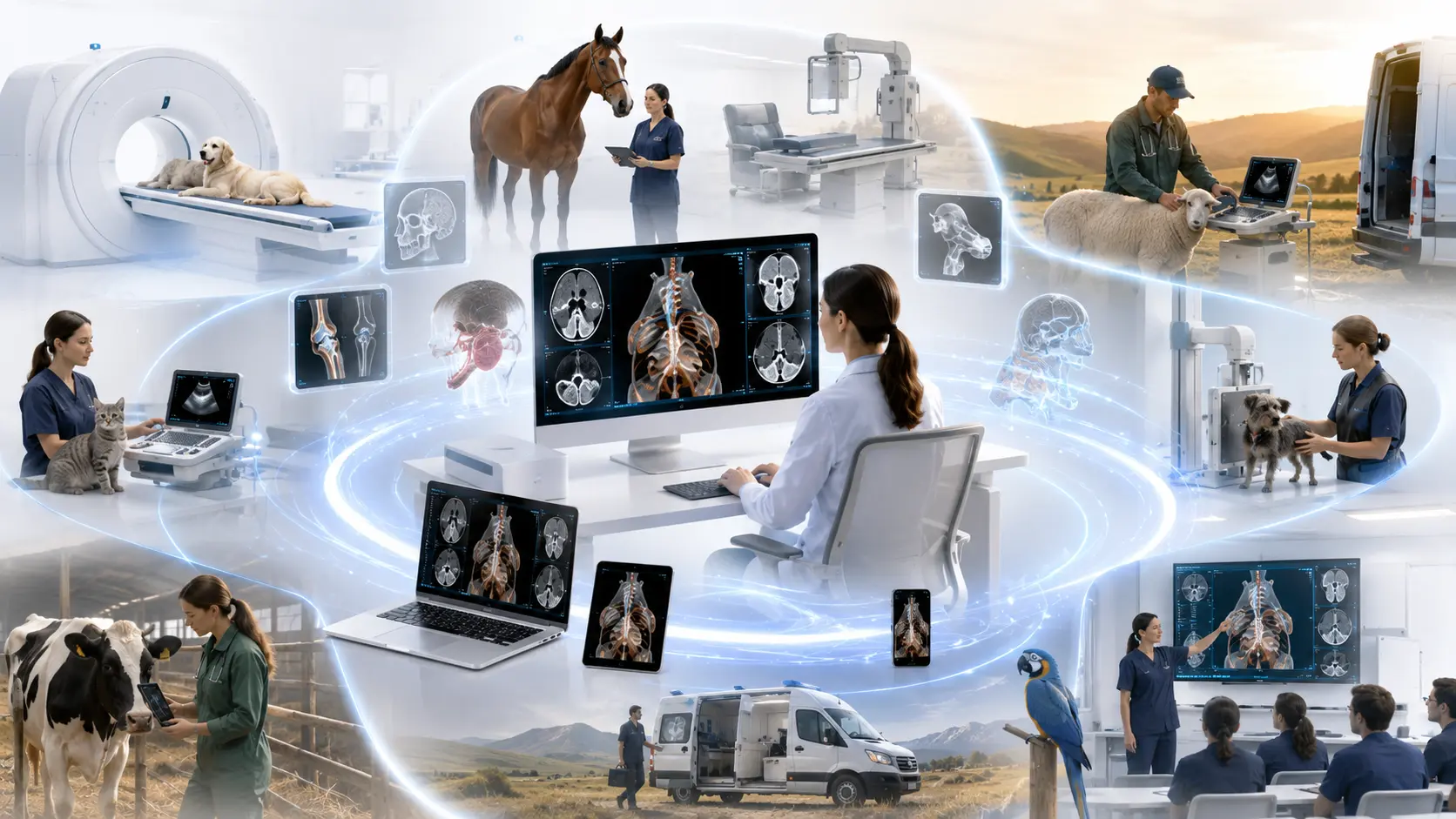
Veterinary imaging increasingly takes place across several locations rather than within a single imaging department. Radiographs may be acquired in a first-opinion clinic, portable digital radiography may be performed beside an equine patient, and CT or MR examinations may be produced at a referral centre. The resulting study may then be reviewed by the attending veterinarian, a surgeon and an off-site radiologist, sometimes using different computers and at different times.
This distributed model creates an important but easily overlooked risk: diagnostic information can be lost after an otherwise satisfactory examination has been acquired. The loss may be obvious, such as an omitted series. It may also be subtle: reconstructed images are mixed with the source dataset, prior examinations cannot be found, veterinary patient information is incomplete, or images are exported as screenshots that no longer retain their original study structure.
DICOM is often described as an image file format. That description is incomplete. The standard addresses the communication and management of medical imaging information, including exchange between acquisition devices, workstations, archives and other systems. Its scope explicitly includes veterinary environments.[1][2]
Within DICOM, an examination is organised hierarchically. A study may contain several series, and each series may contain one or more image or non-image objects. This organisation preserves relationships that would be difficult to reconstruct reliably from a directory of JPEG or PNG files.
- a localiser series;
- one or more axial acquisition series;
- reconstructed series using different slice thicknesses or reconstruction algorithms;
- multiplanar or three-dimensional derived images.
These are not interchangeable collections. They represent different parts or representations of the examination. The American College of Veterinary Radiology recommends, for example, that CT and MR localisers be placed in a separate series, contiguous axial images form a series, and reconstructed datasets be organised separately. Its guidance also defines appropriate study and series organisation for radiography and ultrasound.[3]
A well-designed viewer should make this structure visible. The clinician should be able to understand what was acquired, identify which series is being reviewed and determine whether the examination appears complete.
DICOM supports veterinary information that ordinary image exports may discard
Veterinary imaging has identification requirements that are not always represented well when systems have been adapted from human healthcare. The DICOM patient model includes veterinary-specific attributes for information such as species, breed, neuter status, responsible person and responsible organisation.[5]
These fields can help distinguish the animal patient from the owner or responsible party and preserve relevant context as images move between systems. This does not mean every veterinary system populates every field correctly. DICOM facilitates structured representation, but implementation quality still matters.
A product claiming DICOM support may implement only the services and objects relevant to its purpose. A DICOM conformance statement is therefore useful when comparing systems because it documents which parts of the standard a product supports, although it does not by itself guarantee interoperability.
This distinction is important. “DICOM compatible” is not a complete description of a veterinary imaging workflow. Clinics also need to consider how studies are created, identified, transmitted, retrieved and displayed.
Displaying an image is not the same as reviewing a study
A basic image application can open a picture. Diagnostic review requires more. Window and level adjustment, stack navigation, zoom, pan, measurement and series comparison allow the clinician to interrogate acquired information while preserving its relationship to the complete examination.
For cross-sectional imaging, multiplanar reconstruction provides another example. An axial CT series may be reformatted into sagittal, dorsal or oblique planes to follow anatomical structures that do not align conveniently with the original acquisition plane. The reconstruction does not create new diagnostic information; it presents the volumetric dataset from a different orientation.
Similarly, three-dimensional rendering can help communicate complex spatial relationships to surgeons, referring clinicians, students or owners. It should generally be treated as a complementary representation rather than a replacement for reviewing the source series.
The distinction matters because attractive derived images can give the impression that the entire study has been reviewed when important findings may remain visible only in the original data.
Study organisation becomes part of teleradiology quality
The consequences of incomplete or poorly organised studies become particularly apparent when examinations are submitted for specialist interpretation. A radiologist may need:
- all diagnostically relevant images and series;
- appropriate patient and study identification;
- the clinical history and reason for referral;
- previous examinations for comparison;
- confirmation that the submitted examination is complete.
The ACVR and ECVDI consensus statement on veterinary teleradiology addresses acquisition, transmission, submission quality, quality control and documentation. Its central implication is that interpretation quality depends on more than the competence of the reporting radiologist. Deficiencies elsewhere in the submission chain can affect the standard of care.[4]
A viewer cannot compensate for poor positioning, inadequate exposure or an incomplete examination. Nor can it supply clinical information that was never submitted. It can, however, make omissions easier to recognise by preserving the study hierarchy and presenting series in a coherent form.
This is why the viewer should be considered part of the quality-control environment rather than a passive final step.
The challenge for smaller clinics is continuity
Large referral hospitals may operate dedicated archives, specialist workstations and managed imaging networks. Smaller veterinary clinics often have a different problem.
A radiography system may store studies locally on its acquisition workstation. CT images may arrive from an external referral centre. Historical studies may exist on removable media or another computer. The veterinarian reviewing the case may be in an examination room, at another practice location or working from home during an after-hours consultation.
In such settings, the central challenge is often not advanced visualisation. It is maintaining continuity of access without fragmenting the study.
Cloud-based access can help, but “available in the cloud” is not by itself a measure of clinical quality. The useful question is whether the cloud workflow preserves the original DICOM objects, study hierarchy, metadata and prior examinations while providing appropriate access to authorised users.
The objective is not simply to make an image viewable anywhere. It is to make the complete study reviewable wherever a clinical decision needs to be made.
Applying these principles within Falcon Suite
Falcon Suite separates three related but distinct workflow functions: diagnostic review, study availability and connection to local imaging sources. Falcon Mx provides the local DICOM review environment. Its role is to support study and series navigation, display adjustment, measurements, comparison and multiplanar review. In this model, the software is not merely opening individual image files; it is presenting the examination as an organised clinical study.
Falcon Cloud addresses study availability. It is intended to make current and previous examinations accessible across authorised locations and devices, particularly where a clinic does not operate a full-scale imaging archive. Falcon Gateway connects compatible local imaging sources to the cloud workflow, creating a route between acquisition and wider study availability without requiring the acquisition workstation to remain the only practical point of access.
The value of this arrangement is the separation of responsibilities:
- the acquisition system produces the examination;
- Falcon Gateway supports movement from local imaging sources;
- Falcon Cloud maintains availability;
- Falcon Mx provides the diagnostic interaction layer.
No component replaces appropriate acquisition protocols, clinical history, quality control or specialist interpretation. Instead, the architecture is designed to reduce the likelihood that a complete imaging study becomes isolated on one workstation or is reduced to a set of disconnected exported images.
Six questions to ask when evaluating a veterinary DICOM workflow
Regardless of vendor, a clinic can use the following questions when assessing an imaging workflow:
- Are the original DICOM objects retained? Exported screenshots may be useful for presentations or owner communication, but they should not become the only surviving form of the examination.
- Can users see the complete study and its series structure? It should be possible to distinguish acquisition series, localisers and reconstructed datasets.
- Can current and previous studies be compared efficiently? Access to prior examinations may be important for assessing progression or treatment response.
- Are the necessary review tools available at the point of clinical decision-making? The required capabilities will differ between radiography, ultrasound, CT and MR.
- Can the study be transferred for consultation without being manually reconstructed? A specialist should receive an organised examination rather than an arbitrary selection of exported images.
- Does the vendor publish a DICOM conformance statement? A conformance statement does not prove complete interoperability, but it provides a structured basis for understanding and comparing supported DICOM functions.
These questions are more useful than asking whether a product can simply “open DICOM files.”
Conclusion
Diagnostic imaging quality does not end when the acquisition is complete. It continues through study organisation, transfer, retrieval, display, consultation and comparison with previous examinations.
For veterinary clinics, the practical objective is therefore not merely image access. It is the preservation of diagnostic context across every place where the study may be used.
A capable diagnostic viewer, supported by an appropriate local or cloud-connected workflow, cannot replace clinical expertise. It can help ensure that expertise is applied to the complete study rather than to an incomplete representation of it.
References
Sources cited in this article
- [1]About DICOM: Overview
DICOM Standard Committee / NEMA
DICOM overview and role in medical imaging interoperability.
- [2]ISO 12052:2026 - Health informatics: DICOM including workflow and data management
International Organization for Standardization
DICOM scope, including clinical, research, veterinary, and other medical environments.
- [3]Radiology: Image Resolution & DICOM Guidelines
American College of Veterinary Radiology
Veterinary profession guidelines for image resolution and DICOM study/series organization.
- [4]ACVR and ECVDI consensus statement for teleradiology
Veterinary Radiology & Ultrasound
Best-practice guidance for quality and safety in veterinary teleradiology.
- [5]DICOM PS3.3: Patient Demographic Module
DICOM Standard Committee / NEMA
Current DICOM patient information attributes, including veterinary-relevant species, breed, responsible person and responsible organization fields.